
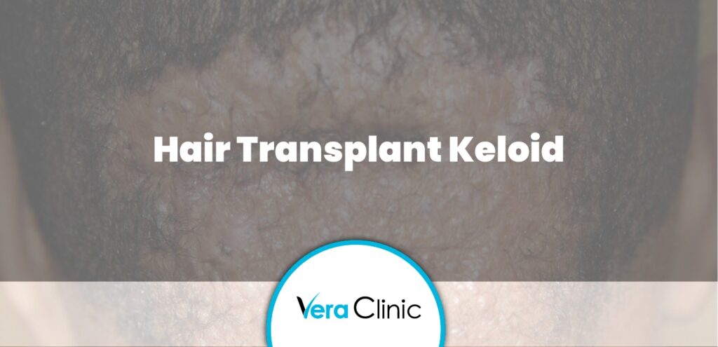

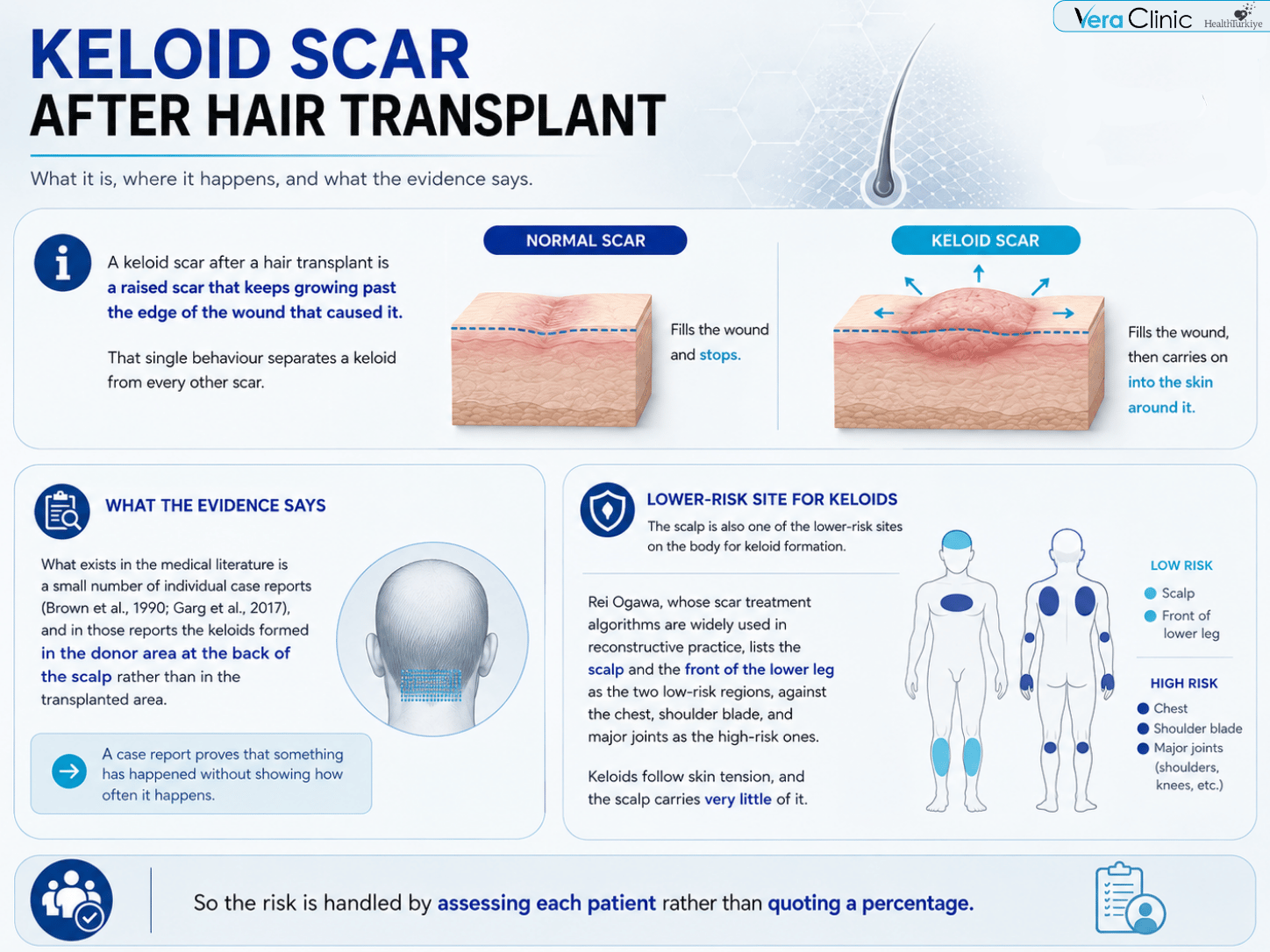
A keloid scar after a hair transplant is a raised scar that keeps growing past the edge of the wound that caused it. That single behaviour separates a keloid from every other scar. A normal scar fills the wound and stops. A keloid fills the wound, then carries on into the skin around it.
Keloid formation after a hair transplant has no published incidence rate, because nobody has measured one. What exists in the medical literature is a small number of individual case reports (Brown et al., 1990; Garg et al., 2017), and in those reports the keloids formed in the donor area at the back of the scalp rather than in the transplanted area. A case report proves that something has happened without showing how often it happens, so the risk is handled by assessing each patient rather than by quoting a percentage.
The scalp is also one of the lower-risk sites on the body for keloid formation. Rei Ogawa, whose scar treatment algorithms are widely used in reconstructive practice, lists the scalp and the front of the lower leg as the two low-risk regions, against the chest, shoulder blade, and major joints as the high-risk ones. Keloids follow skin tension, and the scalp carries very little of it, which is one reason keloid risk is treated as a distinct, closely assessed layer within the broader hair transplant scar picture rather than as a routine expectation.
Key Points
- A keloid keeps growing past the original wound edge, while a hypertrophic scar stays within it; this is the decisive diagnostic feature, confirmed by a tongue-shaped advancing edge visible under the microscope in keloid tissue (Limandjaja et al., 2021).
- No published study has measured the incidence of keloid scarring after hair transplant. The literature contains only individual case reports (Brown et al., 1990; Garg et al., 2017), and in each case the keloid formed in the donor area rather than the transplanted area.
- The scalp is one of the two lowest-risk body sites for keloid formation based on skin tension, per Ogawa’s scar treatment algorithm; high-tension sites such as the chest and shoulder blade carry the greatest risk.
- Genetic variation accounts for about 34% of keloid risk in people of African ancestry versus about 6% in people of European ancestry, and diagnosed keloid prevalence runs at roughly 2% versus 0.4% between the same two groups (Greene et al., 2025; Ung et al., 2023).
- The strip technique (FUT) carries a higher donor-area scarring risk than FUE, since it creates one larger wound closed under continuous tension rather than many small, low-tension extraction points.
- Surgical removal alone has a reported recurrence rate of 45% to 100% depending on site and technique, which is why keloid excision is combined with other treatments rather than performed on its own (Ogawa, 2022).
- Across a Vera Clinic Academy cohort of 420 screened patients, only 11 (2.6%) were excluded at intake for a known personal history of keloid scarring, and most keloid-related disclosures involve family history alone rather than a personal keloid.
- Acne keloidalis nuchae, despite its name, is not a true keloid; it is a distinct inflammatory condition caused by curled hair regrowing into the skin, most common in men of African-Caribbean descent.
What Is the Difference Between a Keloid and a Hypertrophic Scar?
A keloid and a hypertrophic scar differ on four features, of which one is decisive. Both are raised scars, both come from excess collagen, and both are more common in some families than others. The two behave differently after that, and the table below sets the four differences side by side.
| Feature | Hypertrophic Scar | Keloid |
|---|---|---|
| Growth pattern | Stays inside the wound edge, or just at it | Grows beyond the wound edge |
| Time to appear | 4 to 8 weeks after the wound closes | 3 months to several years afterwards |
| Course over time | Thickens for 6 to 8 months, then flattens | Does not flatten and does not mature |
| After removal | Rarely returns once treated | Returns often |
Growth beyond the wound edge is the decisive feature, and it is the one a doctor checks first. A raised scar that has stayed within the original wound is a hypertrophic scar however thick it is, and a raised scar that has crossed into untouched skin is a keloid however thin it is. Size does not decide it and neither does colour.
Limandjaja and colleagues reviewed the evidence for treating the two as separate conditions (Experimental Dermatology, 2021) and found a second marker under the microscope. A keloid shows a distinctive tongue-shaped advancing edge beneath the skin in effectively every case examined, and hypertrophic scars showed it in none of them. Their conclusion was that most of the differences between the two are differences of degree, and that the difference in behaviour is clear enough that the two are worth treating as separate problems.
Getting the distinction right changes what a patient is told. A hypertrophic scar is a healing pattern that resolves. A keloid is an ongoing condition that needs treatment to stop it enlarging, so a scar called the wrong name is a scar managed on the wrong timetable.
Does a Hypertrophic Scar Fade on Its Own?
Yes, a hypertrophic scar fades on its own. It reaches its thickest point within about 6 to 8 months, then flattens and pales over the following months and years. Its inflammatory activity falls away by roughly the three-year mark, which is when the scar stops behaving like an active one.
A keloid follows none of that curve. Keloid tissue shows no change with scar age at all, while hypertrophic scars lose their inflammatory infiltrate as they get older (Limandjaja and colleagues, 2021). Age alone improves one scar and does nothing to the other.
Treatment therefore aims at two different outcomes. A hypertrophic scar is supported while it settles, since the direction of travel is already correct. A keloid is treated to halt its growth, and the earlier that treatment begins, the smaller the intervention needs to be.
How Does a Keloid Scar Form?
A keloid forms when the inflammation that repairs a wound fails to switch off. Healing normally runs through four stages and finishes in the fourth. Bleeding stops, inflammation clears the wound, new tissue fills it, and the scar then remodels and settles. A keloid gets stuck between the second and third stages, so tissue keeps being built long after the wound has closed.
The signal driving that build-up is a growth factor called transforming growth factor beta 1 (TGF-β1), which tells skin cells to make collagen. Keloid skin carries more of it than normal skin does. The more interesting fault sits on the other side of the system. Skin also carries a natural brake on that signal, a protein called Smad7, and keloid fibroblasts show reduced Smad7 activity compared to healthy skin, weakening that brake. The problem is a missing off switch rather than a loud on switch.
The scar then reinforces itself, which is what makes a keloid permanent rather than merely slow. Cells inside a keloid respond to stiffness in the surrounding tissue by producing more collagen, which stiffens the tissue further, which activates them again. Single-cell studies of keloid tissue found that the largest cell population in a keloid is the group tuned to sense mechanical force (Zhang et al., 2025). A scar that manufactures its own activating signal has no reason to stop, and that is the mechanism behind a keloid that never flattens.
Why Does Skin Tension Change Where Keloids Appear?
Skin tension decides which parts of the body form keloids and which parts almost never do. The cells inside a scar read mechanical force directly, so a wound in skin that is pulled and stretched by ordinary movement receives a constant activating signal, while a wound in slack skin does not.
The map of keloid sites follows that rule closely. The chest, the shoulder blade, the shoulders, and the skin over major joints all carry high tension at rest and in movement, and all four are high-risk sites. The scalp sits at the other end, since scalp skin moves very little during normal activity. Low tension is the reason the scalp is a low-risk site, which matters for anyone weighing a scalp procedure against their scarring history elsewhere on the body.
The earlobe is the standing exception, and it is worth naming rather than explaining away. Earlobes carry almost no tension yet appear as the single commonest keloid site in hospital series, accounting for 30.7% of cases in a 2025 Ugandan study of 231 patients (Mbiine et al., 2025). Exposure rather than tension is the likely reason, since ear piercing is the most common keloid trigger in the world.
Why Is Ancestry a Stronger Predictor Than Skin Tone?
Ancestry predicts keloid risk because the risk is inherited, and skin tone is a rough marker for ancestry rather than a cause. The largest genetic study of keloids to date, spanning 7,837 patients and more than 1.5 million controls (Greene et al., Nature Communications, 2025), identified 26 genome regions linked to keloids, of which 12 were confirmed in a separate independent dataset; the table below sets out how far apart the ancestry groups sit on the strongest of those signals.
| Measure | African Ancestry | European Ancestry | East Asian Ancestry |
|---|---|---|---|
| Genetic heritability of keloid risk | ~34% | ~6% | — |
| ITGA11 risk-variant frequency | 35% | 15% | 4% |
| Diagnosed keloid prevalence | ~2% | ~0.4% | — |
The ITGA11 variant, sitting beside a gene that builds a collagen receptor, was the single strongest signal in the African-ancestry analysis, and the diagnosed-prevalence gap lines up with it closely across two independent datasets that agree with each other (Ung et al., JAMA Dermatology, 2023; Greene et al., 2025). Higher risk in this group is real and measurable, and it is a group-level probability rather than a prediction about any one patient; a family history of keloids and existing keloid scars elsewhere on the body are both stronger signals for an individual than skin tone is.
Which Hair Transplant Technique Carries the Higher Scarring Risk?
The strip technique carries the higher scarring risk, because it makes a larger single wound under more tension. Follicular unit extraction, or FUE, removes grafts one at a time through openings under a millimetre wide and leaves small round marks. Follicular unit transplantation, or FUT, removes a strip of skin from the back of the scalp and closes the gap, which leaves one continuous line held together under tension.
Wound size and wound tension are the two things that matter, and the strip method raises both. A closed strip wound pulls at its own edges for as long as it takes to heal, and tension is the mechanical signal that keeps scar-building cells active. Raised and keloidal scarring in the donor area is reported with the strip method more often than with extraction for that reason, one of several risks that factor into why Vera Clinic does not offer FUT hair transplant.
Instrument size works in the same direction within FUE itself. Extraction for Afro-textured hair uses a punch of 0.9 to 1.0 mm in male patients and 0.8 mm in female patients, both inside the 0.8 to 1.2 mm range that defines FUE punches generally, with the exact diameter tested on a few grafts in the operating room before it is committed to (Vera Clinic Academy Database, 2026). A smaller opening is a smaller wound, and Garg et al. (2017) recommended the smallest workable punch on exactly that reasoning after reporting extensive donor-site keloids in an FUE patient.
How Is Keloid Risk Assessed Before Surgery?
Keloid risk is assessed by history and physical examination at consultation. Three things are looked at, and none of them requires a test procedure. The patient’s own account of how previous wounds healed comes first. Existing scars elsewhere on the body are examined directly, since piercings, acne, vaccinations, and past surgery all leave evidence. Family history is taken last, because keloids run in families.
Examination carries most of the weight because of when keloids first appear. Keloid tendency shows itself between the ages of 10 and 30 in most people who have it, so an adult with a keloid tendency has already produced a keloid somewhere. A scar that would appear has generally appeared already, which is why assessment looks at the body a patient already has rather than at a test extraction.
Assessment is the best available tool rather than a guarantee, and the published cases say so directly. One reported patient had no keloids anywhere else on his body and no family history of them, which led his doctors to write that the case confirms how difficult keloid formation is to predict. At Vera Clinic, this history is checked directly at the pre-operative screening, and a personal history of keloid scarring is a reason for rejection in most cases. Across a Vera Clinic Academy cohort of 420 screened patients, 11 (2.6%) were excluded at intake specifically for a known personal history of keloid scarring; most disclosures at consultation involve family history without a personal keloid, and proceed to individual assessment rather than automatic exclusion, a pattern detailed in the Hair Curl and Pigment Graft Survival Study.
“When a patient discloses a family history of keloids, we look at their own skin first. If there’s no personal history of abnormal scarring by the time they’re old enough for it to have shown up, that’s reassuring, but we still note the family history and watch the donor area a little more closely during healing.”
— Dr. Emin Gül, Medical Doctor, Vera Clinic
Is Acne Keloidalis Nuchae a Keloid?
No, acne keloidalis nuchae is not a keloid, despite the name. It is a long-running inflammation of the hair follicles at the nape of the neck that heals with scarring, and dermatology references state plainly that the scars it leaves are not true keloid scars.
The mechanism is different from a keloid’s, and understanding it explains why the condition sits where it does. Tightly curled hair can grow back into the skin instead of clearing it. The trapped shaft behaves as a foreign body, the follicle becomes inflamed, and the inflammation heals with fibrous tissue that traps further hairs. Close shaving, frequent short haircuts, and friction from collars and headgear all add to it. It affects men about twenty times more often than women and is most common in men of African-Caribbean descent, which places it inside the donor zone for exactly the patients this procedure is designed for. Its bearing on donor area planning is covered as part of the Afro hair transplant procedure itself.
What Happens When a Raised Scar Appears After Surgery?
Treatment begins at the first visible sign rather than after a scar has formed. Redness and raised tissue at a healing site are the trigger for treatment, and intralesional triamcinolone acetonide is injected into the site at that point. The same response applies to a hypertrophic scar as to a keloid, so the distinction between the two does not delay treatment.
Acting early is what keeps the treatment small, and there is a mechanical reason for it rather than only a practical one. An established keloid reaches a state where its cells continue producing collagen even without the original signal driving them, which is the best current explanation for why mature keloids resist treatments that would have worked on the same scar months earlier. A raised area caught in its first weeks is a smaller problem than the same area caught in its second year.
How Does a History of Keloid Scars Affect Candidacy?
A history of keloid scars narrows candidacy considerably, and it is assessed rather than assumed. A patient who already has keloid scars is declined. A patient with a history of hypertrophic scarring may also be declined, on the reasoning that a scarring tendency severe enough to matter has generally already shown itself by adult age.
Declining on these grounds is uncommon in practice, because the presentation itself is uncommon. Higher population risk in people of African ancestry is a reason to examine carefully at consultation rather than a reason to expect a keloid, and the two are different things. Ancestry raises the prior probability. Examination of the actual scars a patient carries is what settles it.
Frequently Asked Questions
No, a hair transplant does not cause keloid scars in most patients. Keloid formation after the procedure has no measured rate, and the medical literature records it only as individual case reports (Brown et al., 1990; Garg et al., 2017). Keloids form in people who carry the tendency, so the surgery is the trigger rather than the cause.
Keloid scars after a hair transplant form in the donor area at the back of the scalp in the reported cases, rather than in the transplanted area at the front. Extraction openings sit in the occipital scalp, and that is where the reported scars appeared. The recipient area receives grafts into channels rather than losing tissue.
A keloid appears 3 months to several years after the wound closes, which is later than a hypertrophic scar. Redness and raised tissue at a healing site are the earliest visible signs. Treatment starts at that point rather than waiting for the scar to develop, because an early raised area responds better than an established one.
Black patients carry a higher keloid risk in general, and no study has measured whether that risk carries into hair transplantation. Keloid diagnoses run at roughly 2% in people of African ancestry against roughly 0.4% in people of European ancestry (Ung et al., JAMA Dermatology, 2023). Population risk and surgical risk are two different measurements, and only the first has been taken.
Yes, itching and pain are the two commonest keloid symptoms. Both are reported more often than any other complaint in keloid patients (Greene et al., Nature Communications, 2025). Itching comes from inflammatory activity inside the scar rather than from the skin surface, which is why scratching does not settle it.
A keloid scar is removed surgically and returns often when surgery is the only treatment. Recurrence after excision alone is reported between 45% and 100% depending on the site and the technique used (Ogawa, 2022). Removal is therefore combined with other treatments rather than performed on its own.
- Brown MD, Johnson T, Swanson NA. Extensive keloids following hair transplantation. J Dermatol Surg Oncol. 1990;16(9):867-869.
- Garg S, Kumar A, Tuknayat A, Thami GP. Extensive donor site keloids in follicular unit extraction hair transplantation. Int J Trichology. 2017;9(3):127-129.
- Greene, C.A. et al. (2025). Multi-ancestry meta-analysis of keloids. Nature Communications, 16, 7770.
- Limandjaja, G.C. et al. (2021). The Keloid Disorder. Experimental Dermatology.
- Mbiine, R. et al. (2025). Description of clinical and histopathological characteristics of keloids in a sub-Saharan African population. Skin Health and Disease, 5(5), 326–343.
- Ogawa, R. (2022). The most current algorithms for the treatment and prevention of hypertrophic scars and keloids. Plastic and Reconstructive Surgery, 149(1), 79e–94e.
- Ung, C.Y. et al. (2023). Comorbidities of keloid and hypertrophic scars among UK Biobank participants. JAMA Dermatology, 159, 172–181.
- Vera Clinic Academy. (2026). Graft Survival and Transection Rates Across Hair Curl and Pigment Categories in FUE: A Retrospective Cohort Study at Vera Clinic. Internal institutional data. https://www.veraclinic.net/wp-content/uploads/2026/08/Graft-Survival-and-Transection-Rates-Across-Hair-Curl-and-Pigment-Categories-in-FUE_-A-Retrospective-Cohort-Study-at-Vera-Clinic.docx.pdf
- Zhang, W. et al. (2025). Uncovering a fibroblast differentiation-based keloid classification. Communications Biology, 8, 1387.