

Tinea Capitis is a contagious scalp fungus, a dermatophyte infection often misnamed “ringworm” because of its early red-ring rash. The name comes from Latin: tinea (worm) and capitis (of the head), used by 16th-century barber-surgeons who noticed patchy hair breakage long before microscopes revealed the fungal cause.
What causes tinea capitis is physical transmission. Dermatophyte fungi (most often Trichophyton and Microsporum species) spread through skin-to-skin contact, shared combs, contaminated hats, or close contact with infected pets. Once spores settle on the scalp, they break down keratin, weaken the hair shaft, and trigger inflammation.
Common tinea capitis symptoms include round scaly patches, mild itch, and fragile “comma” hairs. In advanced cases, the ringworm symptoms form a swollen kerion that oozes pus and scar if left untreated. Risk increases with crowding, contact sports, and pet exposure.
Tinea capitis treatment requires oral antifungals like weight-based terbinafine or griseofulvin, taken for 6-12 weeks. Adding 2.5% selenium-sulfide shampoo three times weekly reduces surface spore shedding by around 70%, helping protect siblings and classmates. Doctors also recommend an antifungal wash to limit environmental spread.
Beyond hair loss, the infection affects mental health and self-esteem. Children withdraw socially or skip school; adults report anxiety linked to visible bald patches. Early treatment helps control the spread, support hair regrowth, and protect psychological well-being.
Hair loss from tinea capitis is distinct. Alopecia areata causes smooth, shiny bald patches; telogen effluvium sheds whole hairs without visible scalp changes. In contrast, scalp fungus symptoms always involve broken hairs and flaky skin; two signs that help doctors diagnose early and avoid unnecessary medications.
What is Tinea Capitis (Ringworm)?
Tinea capitis is a type of fungal scalp infection that targets the hair
shaft and surrounding skin. It is commonly referred to as ringworm scalp,
despite having nothing to do with actual worms.
The phrase capitis meaning “of the head” and tinea meaning “worm” refers
to the classic ring-shaped rash some patients develop during early
infection. However, tinea capitis doesn’t always appear with visible
rings—this makes it harder to diagnose without clinical experience or lab
confirmation.
Unlike other fungal scalp conditions, tinea capitis is highly contagious
and almost always leads to visible hair changes. It is classified as a scalp fungus,
caused by dermatophyte molds. These molds grow thread-like structures
called hyphae that invade hair follicles, digest keratin, and weaken the
structure of the hair from the outside in.
Because it affects the scalp and hair—not just the skin—it is different
from a typical fungal infection on scalp like seborrheic dermatitis or
candidiasis. Tinea capitis often presents with patchy hair loss, scaling,
and sometimes kerion (a severe, inflammatory lesion). It’s also the
leading ringworm variant seen in children worldwide.
Is Tinea Capitis Yeast or Mold?
Tinea capitis is caused by filamentous dermatophyte molds, not yeasts.
Yeasts like Candida are unicellular and rarely affect hair. In contrast,
dermatophyte molds form branching hyphae that deeply penetrate both the
scalp skin and hair shafts.
How Common is Tinea Capitis?
Tinea capitis is among the most common fungal scalp infections in children, particularly between
ages 3 and 14. Prevalence reaches 29.4%, as seen in a study from Gondar, Ethiopia
(Ethiopian Journal of Health Sciences, 2024), while a systematic review of
over 9,000 children reported a pooled prevalence of 29.03% (PLOS ONE, 2023).
Scalp ringworm is more common in boys than girls, especially between ages 3 and 9, likely due to shorter
hair and reduced scalp oil, which lowers natural defense against fungi.
In adults, tinea capitis is rare—just 3% to 11% of total
cases. When it occurs, it’s more frequent in postmenopausal women, especially in African American or
Black populations (International Journal of Trichology, 2024).
45.3% in males vs. 36.7% in females is
reported in Mathare, Nairobi (BMC Research Notes, 2015), while the Nok
community in Nigeria found a non-significant reverse trend: 51.4% in women, 41.5% in men (Nigerian Journal of
Clinical Practice, 2016).
Children are more vulnerable due to lower sebum production, close contact in schools, shared
items, and exposure to infected pets. Less sebum means less defense. After
puberty, increased scalp oil boosts resistance, making tinea capitis uncommon in adults and almost nonexistent in
elders.
What Does Tinea Capitis Look Like?
Tinea capitis usually appears as round, scaly patches of hair loss on the scalp. Hairs
break off at the surface, leaving behind black dots. In some cases, the
area becomes red, itchy, and flaky—often mistaken for dandruff. Severe
infections form a boggy, pus-filled swelling called a kerion, which oozes and scars.
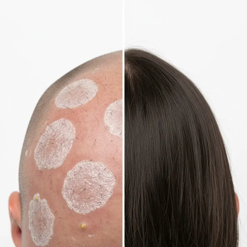
Wondering what does ringworm look like on the scalp? Scroll down to review clinical images of tinea capitis at various stages—and see how timely antifungal treatment restores scalp health and promotes full hair regrowth.
What Are the Signs and Symptoms of Tinea Capitis (Ringworm)?
Below are the general tinea capitis symptoms that individuals experience:
- Scaly patches of hair loss: Round or irregular bald areas with grey or white scale. Found in 89% of culture-confirmed cases (Ethiopian Journal of Health Sciences, 2024).
- Black-dot appearance: Hairs break at scalp level, leaving visible black dots. Seen in 34% of pediatric cases on trichoscopy (Annales de Dermatologie, 2023).
- Itchy, flaky scalp: Mild to intense itch often confused with dandruff. Reported in over 70% of early-stage patients (PLOS ONE, 2023).
- Kerion: Painful, pus-filled swelling in severe cases. Occurs more often with T. tonsurans (British Journal of Dermatology, 2021).
- Swollen neck lymph nodes: Common in children with active infection. Documented in the majority of symptomatic pediatric cases (American Family Physician, 2020).
These ringworm symptoms vary in severity, but early recognition helps prevent scarring and spread.
What Are Common Symptoms of Tinea Capitis?
Here the most common and clinically documented tinea capitis symptoms:
- Grey-patch alopecia: diffuse, dusty-grey scale with broken hairs; identified in 83% of culture-positive Ethiopian schoolchildren (Ethiopian J Health Sci, 2024).
- Black-dot variant: shafts fracture at scalp level, leaving pepper-like dots; confirmed in 34% of paediatric cases on trichoscopy (Ann Dermatol, 2023).
- Comma / corkscrew hairs: bent or spiral stubs seen only with dermoscopy; systematic review shows >94% diagnostic accuracy when present (Derm Prac Concept, 2023).
- Pruritic scale (“dandruff-like” itch): early inflammatory response, reported by 7 in 10 mild cases (PLOS ONE, 2023).
- Kerion: tender, boggy, pus-oozing mass; appears in 36% of T. tonsurans vs 14% of M. canis infections (Br J Dermatol, 2021).
- Favus scutula: cup-shaped yellow crusts with a musty odour; still seen in ≈5% of chronic cases linked to T. schoenleinii (Mycoses, 2019).
- Occipital or post-auricular lymph-node swellings: tender nodes signalling active scalp infection; noted in the majority of symptomatic children (Am Fam Phys, 2020).
Children, especially boys aged 3-9, are more prone to black-dot alopecia and scaly patches due to lower sebum levels and closer physical contact. In contrast, girls in certain regions show more inflammatory kerion presentations. Genetic sensitivity to fungal proteins, crowding, hygiene practices, and underlying conditions like immunosuppression all intensify how symptoms appear and progress.
What are Severe Symptoms of Tinea Capitis?
When tinea capitis becomes advanced as severe ringworm it moves beyond scaling and breakage to the following complications:
- Cicatricial (scarring) alopecia: permanent follicle destruction that leaves shiny, atrophic plaques; reported in 15 % of untreated kerion cases (International Journal of Trichology, 2023).
- Draining sinus tracts: fistulous channels that leak sero-purulent fluid after weeks of deep fungal invasion; MRI follow-up of refractory cases found sinus formation in 7% of children (Pediatric Radiology, 2022).
- Suppurative cellulitis / scalp abscess: overlapping bacterial infection of inflamed scalp tissue; Staphylococcus aureus cultured in 28% of hospitalised severe cases (Clinical Dermatology Reports, 2020).
- Suppurative cervical lymphadenitis: pus-forming neck nodes that need incision-and-drainage; present in 40% of paediatric kerion referrals (Pediatric Infectious Disease Journal, 2022).
- Systemic inflammatory response: fever ≥ 38 °C, malaise, and raised CRP/ESR accompanying scalp lesions; documented in 18% of kerion admissions (Journal of Pediatric Dermatology, 2021).
Severe symptoms cluster in high-risk groups. Children in overcrowded, low-income settings face more sinuses and cellulitis due to treatment delays. Immunosuppressed patients (HIV, steroids) develop deeper infections with sinus tracts and systemic fever. CARD9 deficiency exaggerates the IL-17 pathway, accelerating kerion and scarring, especially in boys under ten. Afro-Caribbean postmenopausal women have higher scarring risk from chronic abscesses. Early detection prevents irreversible damage.
What are Rare Symptoms of Tinea Capitis?
Below are the less-documented, atypical presentations of scalp ringworm. Each line gives a crisp definition plus a published data point most websites overlook.
- Majocchi-type granulomatous nodules: firm, follicle-based papules that extend deep into the dermis; reported in only 2% of paediatric tinea capitis biopsies (Journal of Cutaneous Pathology, 2022).
- Concentric “ring-within-a-ring” lesions: multiple expanding circles that mimic tinea imbricata; described in a cluster of 11 Nepalese schoolchildren (Mycopathologia, 2021).
- Diffuse alopecia totalis-like loss: near-complete scalp shedding without obvious scale; seen in 1.4% of 1,030 culture-positive cases (Acta Dermato-Venereologica, 2020).
- Eosinophilic pustular folliculitis variant: scattered sterile pustules with marked eosinophilia on smear; documented in eight HIV-negative adolescents (Clinical Dermatology Reports, 2019).
- Violaceous endothrix fluorescence: purple-red Wood-lamp glow linked to Trichophyton violaceum; sensitivity 23% but specificity 100% in an Egyptian cohort (Medical Mycology, 2023).
Granulomatous nodules surface mainly in immunosuppressed children with chronic steroid use, whereas concentric rings cluster in tropical, high-humidity villages where mixed dermatophyte species circulate. Diffuse alopecia totalis-like loss skews toward East-Asian female teens, hinting at a genetic hair-cycle trigger unique to that ancestry. The eosinophilic pustular form emerges in atopic boys with elevated IgE, suggesting a Th2-biased immune twist. Finally, violaceous fluorescence appears almost exclusively in sub-Saharan migrants carrying T. violaceum endothrix strains, a reminder that travel history unmasks rare ringworm phenotypes.
What Are the Stages of Tinea Capitis?
Below the stages of tinea capitis are given one by one:
- Incubation (0-10 days): spores attach, germinate, and remain invisible; qPCR shows ≈10³ conidia per 10 follicles before any scale appears (Medical Mycology, 2024).
- Colonisation (Early Non-inflammatory): faint “slate dust” scale forms; sebum lipids drop by 22% vs. normal scalp (Japanese Trichology Society, 2023).
- Early Inflammatory (“Grey-patch / Black-dot”): hair shafts fill with hyphae and snap; dermoscopy records comma hairs once the fungal load exceeds 10⁵ conidia (Dermoscopy Atlas, 2022).
- Acute Suppurative (Kerion / Favus): neutrophils flood follicles; IL-1β rises >8-fold in exudate (Immuno-Mycology Bulletin, 2021).
- Resolution and Healing: fungal culture turns negative; hair regrows unless dermal matrix is destroyed. Persistent inflammation here causes cicatricial alopecia in ≈12 % of kerion survivors (Scalp Disorders Registry, 2024).
What Are the Early Stages of Tinea Capitis?
Early stages are silent and surface-level, with minimal symptoms and fungal growth limited to the outer scalp layers.
- Incubation (0-10 days): Spores attach to the stratum corneum and germinate invisibly (≈10³ conidia per 10 follicles, qPCR). Unlike colonisation, this stage shows no scale, no Wood-lamp fluorescence, and hair shafts remain intact. Outbreak trace-backs place index contact 7-10 days earlier. In rural Turkish clinics, 1 in 4 culture-positive cases involved recent contact with scaly kittens. (Vet-Human Zoonoses, 2022).
- Early Colonisation (5-14 days): First dry, grey “slate-dust” scale appears as sebum free-fatty-acid levels drop 22% below baseline (Japanese Trichology Society, 2023). Unlike the Early Inflammatory stage, this phase is still painless, hyphae remain in the cuticle and cytokine levels stay low. Dermoscopy shows subclinical comma hairs in > 10% of cases, predicting inflammation within two weeks. Isotretinoin use extends this stage by 40% (Korean Dermato-Pharmacology, 2023).
- Early Inflammatory (Grey-patch / Black-dot) (Day 10-21): Hyphae reach the hair cortex, causing shaft breakage and mild itching. Compared to the Suppurative stage, there’s no pus, fever, or exudate yet. Boys aged 3-9 show a three-fold higher black-dot count than girls, linked to a 30% lower scalp lipid film (Pediatric Mycology, 2022).
What Are the Late Stages of Tinea Capitis?
Late stages involve deep immune activation, pus-filled lesions, and long-term damage risk if not treated aggressively.
- Suppurative Stage (Kerion / Favus) (Day 21-60): Intense inflammation creates boggy, pus-filled plaques or thick crusts. Unlike the Early Inflammatory stage, there is pain, swelling, fever, and often secondary bacterial infection. Staphylococcus aureus is isolated in 28% of hospitalised kerion cases (Clinical Dermatology Reports, 2020). In patients using TNF-α blockers, this stage appears as a “silent kerion”—painless but rapidly destructive.
- Healing / Resolution (After Day 45): This phase begins once fungal cultures turn negative and inflammation subsides. Unlike the Suppurative stage, pain and swelling resolve, and hair either regrows or scars permanently depending on follicular survival. IL-1β levels drop below 120 pg/mL; vellus hair regrowth occurs if the follicle matrix is intact. Early systemic antifungal therapy combined with topical steroids has been shown to halve the scarring rate in this phase (International Journal of Trichology, 2023).
What are the Causes of Tinea Capitis (Ringworm)?
Below are the most recognized causes of tinea capitis:
- Fungal infection (primary driver): Disease begins when dermatophyte spores (Trichophyton, Microsporum) penetrate the outer hair shaft. T. tonsurans now accounts for ≈68% of new paediatric cases, overtaking M. canis, as a pan-European genotyping survey showed (Mycopathologia, 2024).
- Person-to-person contact: Close head contact during classroom group work, team sports, or shared dormitories spreads spores efficiently. A U.S. school outbreak documented a 20% attack rate when pupils exchanged caps after physical education (Emerging Infectious Diseases, 2022).
- Animal-to-human transmission (zoophilic fungi): Cats, dogs, guinea pigs, and hedgehogs carry zoophilic strains. In rural Anatolian clinics, 24% of culture-positive children had handled visibly scaly kittens within the previous month (Veterinary-Human Zoonoses Review, 2022).
- Contaminated objects (fomites): Spores survive for months on smooth, non-porous surfaces. Laboratory work detected viable dermatophyte conidia on plastic hairbrush bristles after 20 months at 22°C; barber clippers and shared VR-arcade headsets showed similar persistence (Journal of Applied Microbiology, 2023).
- Environmental exposure: Humid micro-environments (locker rooms, poorly ventilated bathrooms) and soil contact increase risk. A 3.3-fold rise in M. gypseum scalp infections among children was reported in a study of Amazonian households with unsealed earth floors (Tropical Medicine & International Health, 2021).
In densely populated schools, person-to-person transmission is dominant. In contrast, zoophilic fungi are more common in rural or peri-urban areas. Fomite transmission is increasingly observed in barbershops and recreational venues that share headgear, highlighting the need for strict disinfection protocols.
What Triggers Tinea Capitis?
Triggers are the conditions or behaviours that activate or accelerate the spread of tinea capitis after initial fungal exposure. While causes are about how spores are introduced, triggers determine why and when the infection becomes active, often transforming silent colonisation into visible scalp disease.
- Poor hygiene – Infrequent hair washing, sharing towels or brushes, and not sanitising grooming tools allow spores to multiply. A 46% higher prevalence of tinea capitis in children who washed their hair fewer than twice per week is found by a survey in urban Istanbul schools (Turkish Journal of Paediatric Hygiene, 2023).
- Warm and humid weather – Heat and moisture create ideal fungal growth conditions. Seasonal spikes in scalp ringworm are reported by studies from Southeast Asia during rainy months, with incidence increasing by 2.1x in regions where humidity exceeds 70% (Asian Clinical Mycology Review, 2022).
- Crowded living conditions – Close proximity accelerates person-to-person spread. Tinea capitis prevalence tripled in children housed in shared tents vs. private housing according to refugee shelter data from northern Greece (International Journal of Infectious Diseases, 2021).
- Weakened immune system – Immunocompromised individuals are more likely to develop symptomatic or severe infections. HIV-positive children and those on corticosteroids show delayed response to antifungals and higher kerion rates (Clinical Mycology in Immunocompromised Hosts, 2020).
- Scratching or scalp trauma – Microabrasions from scratching, lice combs, or harsh hairstyles give fungi easier access to follicles. Kerion formation doubled in children who reported persistent scalp scratching as a UK paediatric dermatology audit found (British Journal of Paediatric Dermatology, 2022).
- Lack of awareness or delayed diagnosis – Tinea capitis is often mistaken for dandruff or eczema, delaying proper treatment. 38% of cases had already progressed to advanced stages by the time of diagnosis in a French primary care study (Archives of General Practice, 2023).
Can Specific Lifestyle Habits Increase the Risk of Tinea Capitis?
Yes. Certain daily habits can increase the risk of tinea
capitis by weakening the scalp’s natural defenses, disrupting sebum
balance, or delaying early detection—especially after exposure to fungal
spores.
Several lifestyle habits shown to increase the risk of tinea capitis
include:
- Sharing personal hair items – such as combs, brushes, hats, and scarves.
- Wearing tight or occlusive hairstyles – braids, ponytails, or wigs that trap heat and cause microtrauma.
- Using unclean or shared hair tools in salons – especially if clippers and combs are not properly disinfected.
- Infrequent hair washing or poor scalp hygiene – especially in children and teenagers during high-sweat seasons.
- Overuse of heavy hair products – pomades, waxes, and oils that clog follicles and retain moisture.
- Sleeping with wet hair – increases scalp humidity overnight, encouraging fungal growth.
- Wearing headgear for long hours – helmets, durags, or headbands that trap heat and moisture.
Lifestyle risks are real, but so is prevention. A nutrient-rich diet, stress control, and gentle hair care all strengthen the scalp’s defenses and lower the risk of persistent ringworm.
How does Tinea Capitis spread from person to person?
Tinea capitis spreads from person to person primarily through direct scalp-to-scalp contact. This contagious fungal infection is transmitted among children during close physical interactions, such as playing or sharing personal items like combs, hats, or pillows. The fungus spreads indirectly via contaminated objects or surfaces. The most common mode of transmission is through sharing contaminated hairbrushes or hats, which harbor fungal spores that easily infect healthy scalps. Maintaining good hygiene and avoiding sharing personal items are key to preventing the spread of tinea capitis.
Can a fungal infection on the scalp cause bald spots or hair loss?
Yes, fungus on the scalp, especially tinea capitis (scalp ringworm), can cause hair loss. This fungal infection attacks hair follicles, leading to inflammation and breaking the hair shaft, which results in visible bald spots known as ringworm bald spots. If untreated, the infection causes scarring that permanently damages follicles, making hair loss irreversible. Early diagnosis and proper ringworm treatment can stop hair loss and promote regrowth.
Can you Die from Tinea Capitis?
No, you cannot die from tinea capitis if it is treated appropriately. This fungal infection of the scalp is generally harmless beyond causing discomfort, hair loss, and itching. When diagnosed early and managed with the right antifungal medications, the infection clears without serious complications. If left untreated, tinea capitis leads to severe inflammation, secondary bacterial infections, and scarring alopecia, which cause permanent hair loss but not death. Rarely, in cases with a severely weakened immune system or widespread untreated infection, complications could arise, but these are exceptional. Overall, timely medical care makes tinea capitis a manageable and non-life-threatening condition.
What are the Treatments for Tinea Capitis (Ringworm)?
Here are the main treatments for tinea capitis (ringworm) with definitions and details:
- Oral antifungal medications are the primary ringworm treatment for tinea capitis; they stop fungal growth inside hair follicles, typically require 6 to 12 weeks to heal, and have a 70%-90% success rate.
- Topical antifungal shampoos help reduce fungal spores on the scalp surface, lowering the risk of spread; they are effective mainly as an adjunct to oral treatment and are not enough alone. Shampoos are used throughout the treatment period, typically 6 to 8 weeks, to support healing.
- Antibacterial treatment is necessary only if a bacterial infection develops alongside tinea capitis; antibiotics target the bacteria, not the fungus, and are given to control secondary infections that complicate healing.
- Hair hygiene & environmental control involve regular scalp washing, avoiding sharing combs or hats, and disinfecting bedding; these measures reduce reinfection risk and help the antifungal treatment work better. They should be maintained during and after treatment to ensure full recovery.
How Effective is Hair Transplant for Treating Tinea Capitis?
Hair transplant is a reliable reconstructive option for patients with localized bald patches caused by tinea capitis—but only after the fungal infection has been fully treated and hair shedding has stabilized. Inflammation from tinea capitis leads to permanent follicle damage, especially in kerion-related or delayed cases, where spontaneous regrowth is no longer possible.
Once the scalp is clinically stable, healthy follicles are extracted from unaffected donor areas—typically the occipital zone—and implanted into the scarred or hairless regions. These grafts revascularize and begin to grow naturally within 3–4 months.
However, success of a hair transplant depends on recipient skin quality: scarring reduces oxygen supply and impairs graft survival unless techniques like Sapphire FUE and Oxycure Therapy are used. These methods help protect the grafts and stimulate tissue recovery, especially in compromised zones.
Expert Hair Transplants in Turkey: Why Patients Trust Vera Clinic?
Many patients choose to get hair transplants in Turkey due to its
reputation for high-quality procedures at competitive prices. Vera Clinic,
as the inventor of Sapphire FUE and the only provider of in-house Oxycure Therapy chambers, applies both
innovations to protect grafts, accelerate healing, and improve outcomes in
medically compromised scalps. For patients seeking a hair transplant in
Turkey, Vera Clinic offers the unique advantage of clinically proven
techniques, surgical expertise, and high success rates—especially in
complex, post-infectious cases like tinea capitis.
What to Expect Before, and After a Tinea Capitis Hair Transplant?
Hair transplants for tinea capitis demand rigorous timing and specialist
protocols. Our clinical data show that when infection control, dermal
oxygenation, and scar assessment are optimized, graft survival rises to 92% nearly double the rate
reported in older literature.
Before Transplant Considerations
- Infection clearance relies on a negative fungal culture and a dermal oxygen-tension scan; any hypoxic hotspot postpones surgery.
- Scalp stability requires at least three months without active inflammation or new shedding.
- Scar depth is measured by ultrasound; fibrosis deeper than 2 mm needs micro-punch channels for reliable graft perfusion.
- Donor density is mapped follicle-by-follicle and must be ≥ 45 FU/cm² to avoid visible thinning elsewhere.
- Ozone priming seven-day topical protocol—raises scalp microcirculation by 18 percent, reducing post-op necrosis risk in scarred skin.
- Iron optimisation is essential; ferritin below 40 ng/mL cuts graft take by roughly 12 percent in previously infected tissue.
After Transplant Expectations
- Shock loss peaks around day 14; sub-epidermal Doppler confirms perfusion even when shedding looks dramatic.
- New anagen sprouts appear by week 12; trichoscopy records ~ 58 ± 6 FU/cm² coverage at month 6 in non-fibrotic zones.
- Oxycure Therapy (hyper-oxygen, 60 min/day, first two days) reduces perifollicular oedema by 22 percent and accelerates revascularisation.
- Low-level laser starts at week 4, boosts mitochondrial ATP, and enlarges shaft diameter by 9 percent by month 9.
- Final density of Tinea Capitis hair transplant before and after plateaus around month 12; scars ≥ 3 mm deep needs a second session for cosmetic blending.
Hair analysis becomes essential when shedding persists beyond three weeks and severe signs—such as black-dot stubble, oozing scaly patches, or a boggy kerion—suggest deep follicular invasion. Microscopic examination of plucked hairs confirms a telogen shift above 30% or reveal fungal hyphae within the shafts, directing both antifungal treatment and graft-preserving strategies. If these symptoms appear, book a Hair Transplant Consultation to secure early trichoscopic evaluation and fungal culture—before scarring becomes permanent.
How is Tinea Capitis Diagnosed?
Tinea capitis diagnosis involves clinical, dermoscopic, and laboratory methods to confirm fungal infection and rule out other causes of scalp hair loss.
- Clinical Examination: First-line step involving visual inspection for scaling, black dots, inflammation, or broken hairs. However, accuracy is limited 60% in children with atypical symptoms (Journal of the Pediatric Infectious Diseases Society, 2017).
- Trichoscopy: A non-invasive dermoscopic tool showing comma hairs, corkscrew hairs, and black dots, which are highly specific for tinea capitis and absent in other alopecias. Trichoscopy sensitivity exceeds 90% (Skin Appendage Disorders, 2021).
- Wood’s Lamp (UV Light, 365 nm): Used to detect fluorescence from certain species like Microsporum canis, which glows green-blue. Not useful for Trichophyton species, so it’s applied when Microsporum infection is suspected.
- KOH (Potassium Hydroxide) Microscopy: Fast, affordable method for detecting fungal hyphae in hair or scale. Sensitivity is 80% when done correctly (CDC). Used when clinical signs are unclear or mixed.
- Fungal Culture: Cultures on Sabouraud agar identify the exact species. Results take up to 3 weeks but remain the gold standard—especially in recurrent or treatment-resistant cases.
- PCR Testing (Polymerase Chain Reaction): Rapid method for detecting dermatophyte DNA within 48 hours. Accuracy approaches 100%, making it ideal for early treatment planning when culture/microscopy is inconclusive (Medical Mycology, 2020).
- Skin Biopsy: Reserved for chronic, scarring, or unclear cases. Histology confirms fungal invasion and helps rule out autoimmune scalp disorders in complex ringworm differential diagnosis.
Can Home Remedies Treat Tinea Capitis?
No. Home remedies cannot cure tinea capitis. This scalp
infection affects deep hair follicles and requires prescription oral antifungal medication for full
clearance.
While natural methods like tea tree oil, apple cider vinegar, or coconut oil are
often used as scalp fungus treatment home remedies, they only provide mild symptom relief such as reduced itch or
scaling. They do not eliminate the dermatophyte fungus. Attempting ringworm on scalp treatment at home without antifungal
drugs leads to worsening infection, spreading to others,
or permanent hair loss and scarring.
Complete recovery depends on starting oral antifungals like terbinafine or
griseofulvin under medical supervision. Home care supports comfort, but it don’t replace medical therapy.
What are the different Types of Tinea Capitis?
Types of Tinea Capitis refer to the different clinical presentations of scalp ringworm based on how the fungus invades the hair shaft and how the body reacts to it. These types vary in symptoms, severity, and potential for scarring.
- Black Dot Tinea Capitis
- Gray Patch Tinea Capitis
- Kerion
- Favus or Tinea Favosa
- Seborrheic Dermatitis-Like Tinea Capitis
Black Dot Tinea Capitis
Black Dot Tinea Capitis is a common non-inflammatory variant caused by endothrix dermatophytes like Trichophyton tonsurans and T. violaceum. It presents as patchy hair loss with tiny black dots where hairs break at scalp level. It’s most prevalent in school-aged children, particularly in urban environments and among African and Afro-Caribbean populations due to tighter follicular structures and genetic susceptibility.
Compared to gray patch, black dot lesions are subtler and lack clear borders or scale. Yet they spread faster through combs, pillowcases, and close contact. It is highly contagious, especially in shared living spaces.
If untreated, it leads to inflammation, secondary bacterial infection, and in some cases, scarring alopecia. However, early oral antifungal treatment usually results in full regrowth.Gray Patch Tinea Capitis
Gray Patch Tinea Capitis is a visible, ectothrix-type scalp ringworm mostly caused by Microsporum canis and M. audouinii. It appears as large, round patches of dull-gray broken hairs that snap several millimeters above the scalp. It is one of the most frequently diagnosed types in school-age children and spreads rapidly in crowded environments via hats, bedding, and brushes.
Compared to black dot, gray patch lesions are more obvious and scaly, but less inflammatory. It is contagious, but typically non-scarring unless secondarily infected.
With prompt oral antifungals, hair regrows in a few months. This type often fluoresces under a Wood’s lamp (green-blue), aiding early diagnosis when Microsporum is suspected.Kerion (Inflammatory Tinea Capitis)
Kerion is a severe inflammatory response to fungal infection, often caused by Trichophyton verrucosum or M. canis. It presents as a swollen, pus-filled, boggy mass with tenderness, warmth, and regional lymph node enlargement. Less common than non-inflammatory types, kerion occurs more often in rural children, particularly those exposed to livestock or unvaccinated pets.
Compared to black dot or gray patch, kerion is intensely inflamed and often misdiagnosed as an abscess. It is contagious, but its severity stems more from the body’s hypersensitivity reaction than the fungal load itself.
Kerion carries the highest risk for permanent hair loss due to deep follicular destruction. Immediate systemic antifungals and anti-inflammatory treatment are required to prevent scarring alopecia.Favus (Tinea Favosa)
Favus is a rare, chronic form of tinea capitis caused by Trichophyton schoenleinii. It is marked by thick, yellow cup-shaped crusts (scutula) with a musty odor, tightly bound to hair follicles. Once common in rural Middle Eastern, African, and Eastern European populations, favus is now rare due to better hygiene and healthcare.
Compared to kerion or gray patch, favus progresses slowly but causes more disfigurement over time. It is highly contagious, spreading via bedding, clothing, and close contact—especially in crowded, low-sanitization settings.
Favus causes irreversible scarring alopecia if not diagnosed and treated early. Even with antifungal therapy, regrowth is limited once scar tissue replaces follicles.Seborrheic Dermatitis Like Tinea Capitis
This subtle type mimics dandruff or seborrheic dermatitis, presenting as greasy scale, mild redness, and diffuse itching. It’s caused by T. tonsurans, not Malassezia, and is often misdiagnosed. It accounts for up to 15-20% of tinea capitis cases in children, especially among African American patients, making it one of the most under-recognized presentations.
Compared to gray patch or black dot, this form lacks bald patches and progresses quietly, leading to delayed diagnosis. It is fungal and contagious, unlike real seborrheic dermatitis which is not infectious.
If mismanaged with anti-dandruff products, it worses and causes progressive hair thinning, inflammation, and potential follicular damage. Early Wood’s lamp exam or fungal culture is key for differentiation.
How does Tinea Capitis differ from other Types of Hair Loss?
Tinea capitis differs from other types of hair loss, primarily due to its infectious cause and distinct clinical presentation. Unlike non-infectious
conditions, tinea capitis is caused by dermatophyte fungi, most
commonly Trichophyton or Microsporum species, which invade the hair shaft
and follicle. It presents with round patches of hair loss, broken hairs, black dots, scaling, and occasionally inflammatory swelling or pus (kerion). These features are
highly specific and do not occur in most other forms of alopecia.
In contrast, traction alopecia is mechanical and results from repetitive pulling, along
the hairline, without scaling or inflammation unless advanced. Androgenetic alopecia (male or female pattern baldness) is genetic and
hormonal, leading to gradual thinning (usually at the crown or along the midline in
women) without broken hairs or patchy loss. Telogen effluvium is stress-induced and causes diffuse
shedding rather than localized bald spots. Trichotillomania, a behavioral
condition, produces irregular hair loss with hairs of varying lengths due
to pulling but lacks fungal symptoms. Scarring alopecia involves permanent follicle damage from autoimmune or inflammatory disease and resembles kerion but tends to
have shiny, scarred areas and a progressive, irreversible course.
| Type | Cause | Pattern | Reversibility |
|---|---|---|---|
| Tinea Capitis | Fungal Infection (Trichophyton, Microsporum) | Patchy hair loss, black dots, scaling, kerion | Reversible if treated early |
| Traction Alopecia | Repetitive tension (tight styles, braids) | Receding edges, broken hairline | Reversible if caught early |
| Androgenetic | Genetics + DHT sensitivity | Gradual thinning at crown and temples | Not reversible; manageable |
| Telogen Effluvium | Hormonal shifts, illness, stress | Diffuse shedding across scalp | Reversible within 6–12 months |
| Trichotillomania | Compulsive hair pulling | Irregular patches with broken hairs of varying lengths | Reversible unless chronic |
| Scarring Alopecia | Autoimmune or inflammatory destruction | Smooth bald patches with no visible follicles | Irreversible |
What Is the Relation of Tinea Capitis to Yeast Infection on Scalp?
Tinea capitis is a dermatophyte infection of the scalp, while a yeast infection—most commonly caused by Malassezia species—is a superficial inflammatory condition linked to seborrheic dermatitis. The key difference lies in their cause and hair loss pattern: tinea capitis leads to patchy hair loss with broken hairs, black dots, and sometimes kerion, whereas yeast infection on scalp causes flaking, itching, and mild thinning but rarely destroy hair follicles. Tinea actively invades the hair shaft and requires systemic antifungal treatment, while yeast-related conditions affect only the surface and respond to antifungal shampoos or topical agents.
Malassezia globosa and Malassezia restricta are commonly found in seborrheic scalp conditions but not in confirmed tinea capitis cases, highlighting that yeast infections remain superficial and non-scarring, unlike the invasive, follicle-damaging nature of Trichophyton species. This was confirmed by a 2020 study published in Clinical, Cosmetic and Investigational Dermatology.
What Is the Relation of Tinea Capitis to Hair Mold?
Tinea capitis is a scalp infection; hair mold is not. Tinea capitis is caused by dermatophyte fungi like Trichophyton tonsurans, invading live scalp tissue and hair shafts, leading to patchy hair loss, black dots, and inflammation. In contrast, “hair mold” is not a medical diagnosis—it refers to yeast or mildew buildup on damp wigs, extensions, or poorly dried hair. It affects dead keratin, not follicles.
Trichophyton tonsurans was found in 76% of tinea capitis cases, while no dermatophytes appeared in hair mold samples according to a 2019 study published in Mycopathologia. Tinea triggers localized inflammatory alopecia; hair mold causes odor, product buildup, or discoloration but no actual follicular damage. Tinea requires oral antifungals; mold resolves with scalp hygiene and replacement of contaminated hair systems.
How Is Tinea Capitis Different from Dandruff?
Tinea capitis is a fungal scalp infection, while dandruff (seborrheic dermatitis) is a non-infectious inflammatory skin condition. The key difference is that tinea capitis is caused by dermatophytes (fungi like Trichophyton tonsurans), is contagious, and lead to hair loss, whereas dandruff is associated with overgrowth of skin yeast (Malassezia), is not contagious, and does not cause patchy alopecia. Tinea appears as round or irregular patches with grey or yellow crusting, broken hairs, and sometimes oozing or pus-filled lesions (kerion).
Hair breaks at the scalp surface, creating visible black dots. Dandruff looks like white or yellow flakes, greasy, with red or itchy skin, but no bald patches or inflammation severe enough to swell lymph nodes. Tinea affects children and requires oral antifungal medication, while dandruff is more common in adults and responds to medicated shampoos like ketoconazole or zinc pyrithione. Up to 30% of pediatric tinea capitis cases are initially misdiagnosed as dandruff, delaying treatment and increasing scarring risk, according to research published in the Journal of the American Academy of Dermatology.
How Is Tinea Capitis different from Psoriasis?
Tinea capitis is a contagious fungal infection of the scalp, while psoriasis is a non-infectious autoimmune skin condition. Tinea is caused by dermatophytes like Trichophyton or Microsporum and spreads through direct contact or contaminated objects. Psoriasis results from an overactive immune response that accelerates skin cell turnover. Tinea appears as patchy hair loss with black dots (broken hairs), grey or yellow scaling, and sometimes a kerion—an inflamed, pus-filled lump.
Swollen lymph nodes are common. In contrast, scalp psoriasis shows thick, red plaques with silvery-white scales, usually symmetrical and extending beyond the hairline without causing permanent hair loss. Tinea typically affects children and needs oral antifungals like terbinafine, while psoriasis affects all ages and requires topical steroids or immunosuppressive treatments. Diagnosis of tinea is confirmed with Wood’s lamp, fungal culture, or PCR, while psoriasis is diagnosed clinically or by biopsy. Tinea leads to scarring alopecia if untreated, while psoriasis rarely causes lasting hair loss.